av
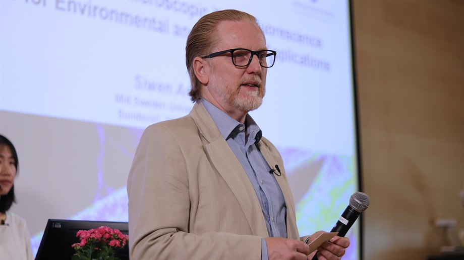
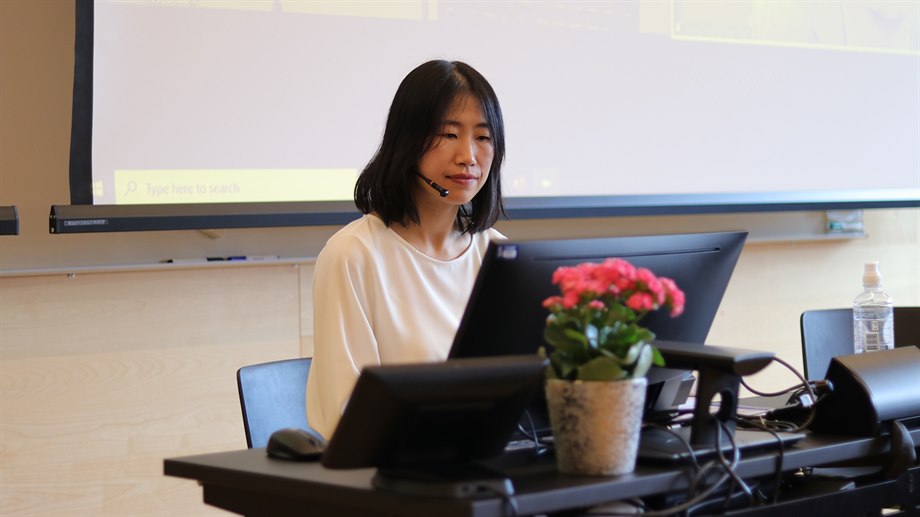
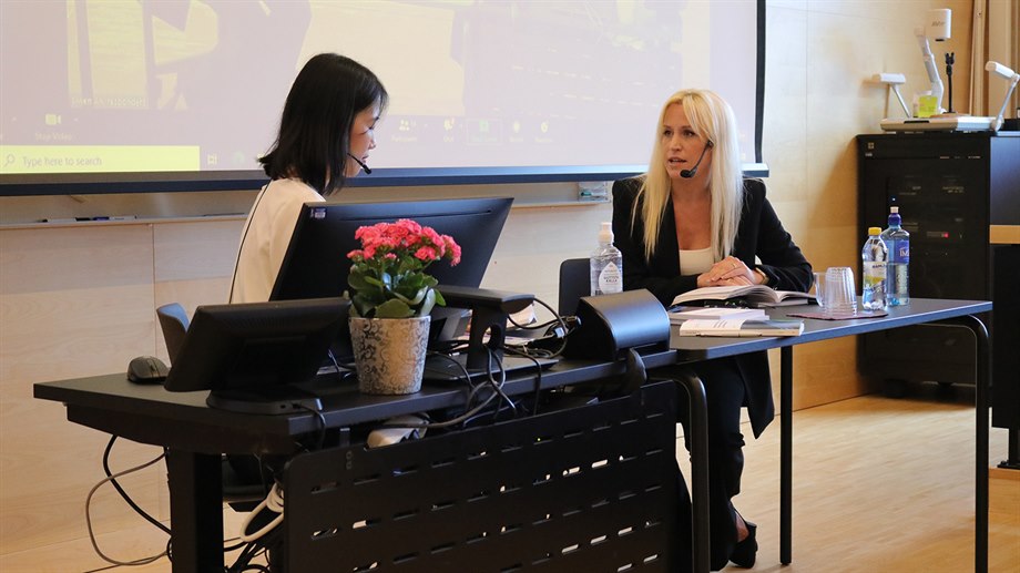
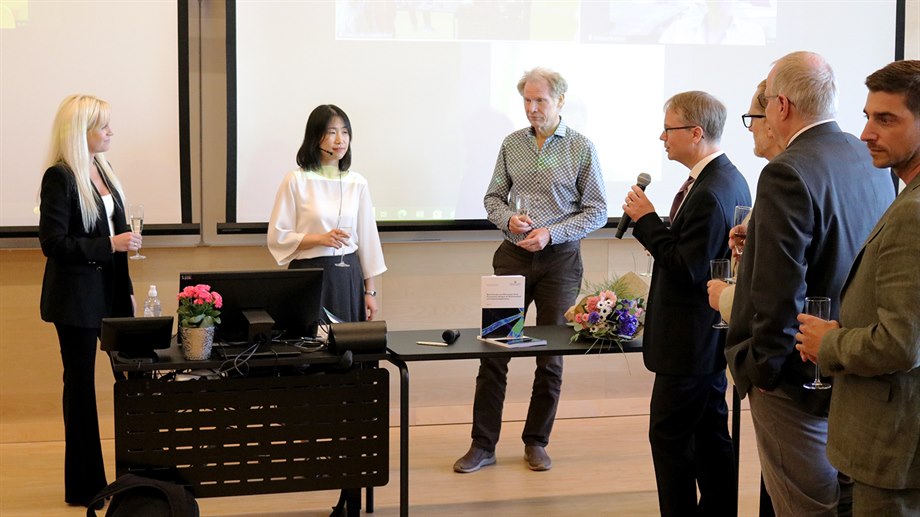
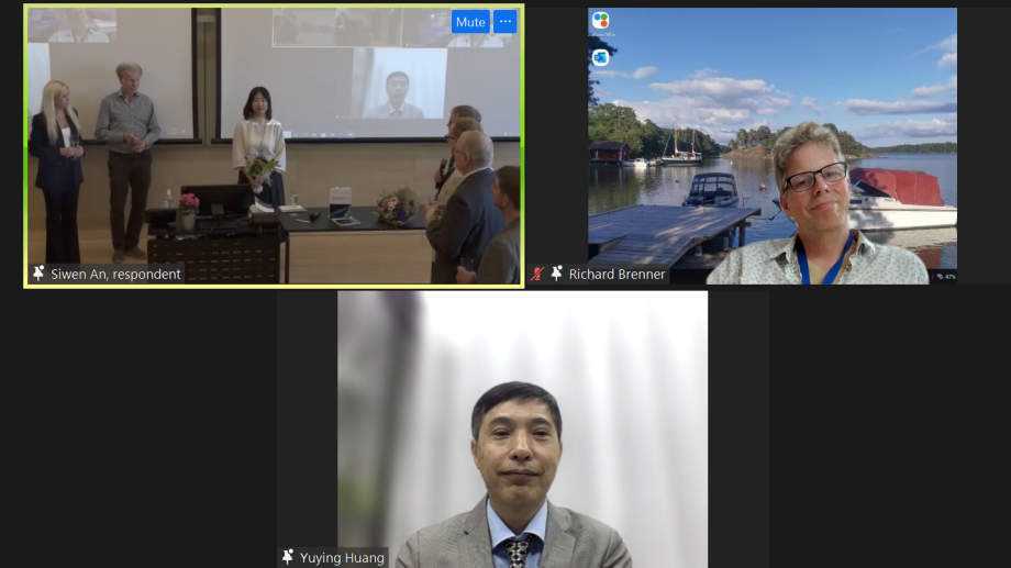
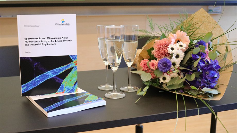
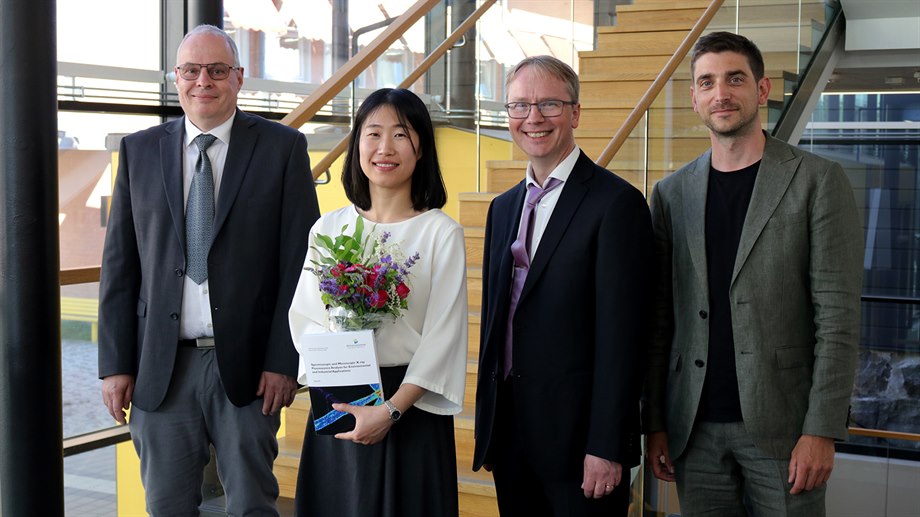
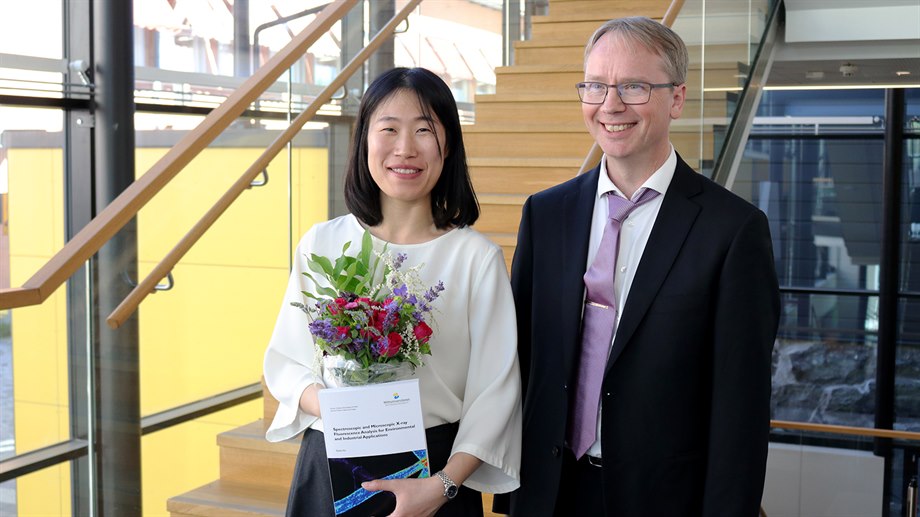
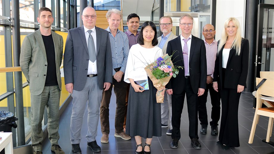
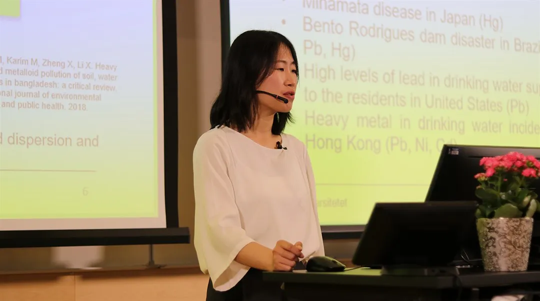
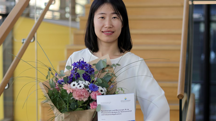
Fredag den 17 juni var det dags för Siwen An att presentera och försvara sin avhandling "Spectroscopic and Microscopic X-ray Fluorescence Analysis for Environmental and Industrial Applications" vilket hon framgångsrikt gjorde.

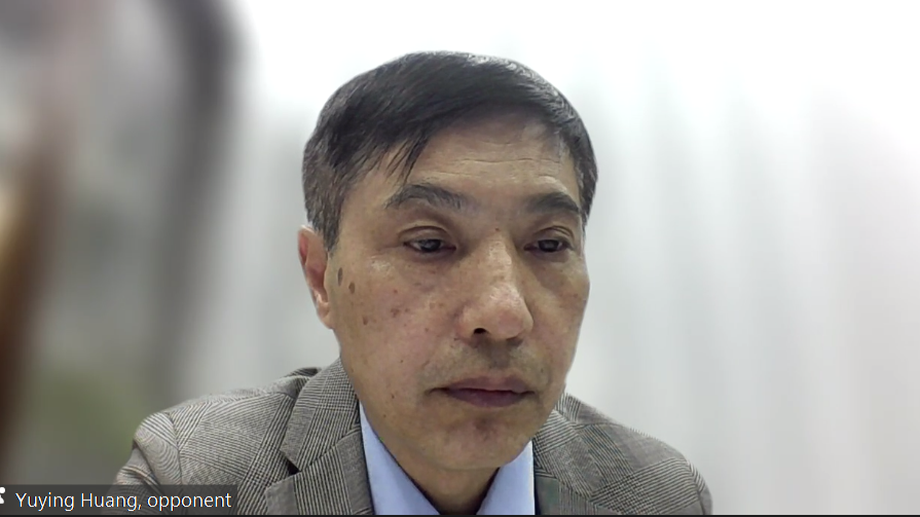
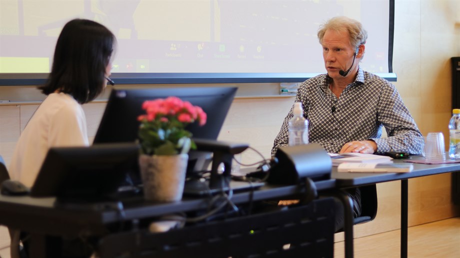
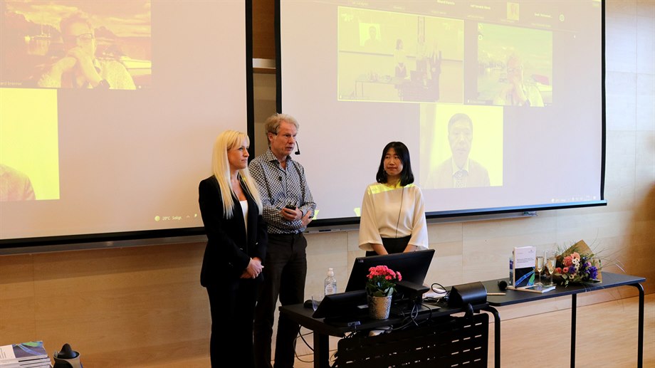
Siwen började med att presentera sin avhandling. Därefter var detdags för opponenten Professor Yuying Huang, Shanghai Institute of Applied Physics, att ställa sina frågor till Siwen. När opponenten var klar tog betygsnämnden över och ställde sina frågor. I betygsnämnden deltog Professor Richard Brenner, Uppsala Universitet, Professor Anders Hallén, KTH Kungliga Tekniksa Högskolan och Dr. Gabriella Josefsson, Hamamatsu Photonics.
Siwen Ans handeldare var Docent Börje Norlin och biträdande handledare var docent Göran Thungström och Dr. David Krapohl, Mittuniversitetet.
Heavy metals are well-known environmental pollutants due to its potential impact on associated ecosystems and human health. Thus, it is important to monitor the levels of heavy metals in the environment. X-ray fluorescence (XRF) analysis is a powerful and effective screening tool in measuring the concentration of multi-elements simultaneously.
This thesis provides insight into development and implementation of XRF instruments for environmental monitoring and industrial process control. The XRF method was compared with a commercial scanning electron microscope with energy dispersive spectroscopy (SEM-EDS) for fly ash samples. Qualitative analysis and semi-quantitative analysis of Na, S, Cl, K and Cd in incineration fly ash were performed with these two similar techniques. One of the challenges of using XRF is the scattering background noise from the primary beam, which decreases the detection limit and the sensitivity of the measurement system. Hence, an X-ray beam filter was chosen to suppress the background noise for a specific element, Cr, in leachate. Numerical simulations and experiments were developed to find the proper filter material and thickness by calculating the X-ray fluorescence intensities and the signal-to-noise ratio. The developed system is capable of online monitoring of Cr levels, to certify that the concentration is below the threshold level in leachate. An XRF prototype was built and calibrated for underwater Hg analysis in maritime wet sediment using a radioisotope source. The presented results show that it is possible to detect Hg by K-shell emission thus enabling XRF analysis for sediment underwater.
For non-homogeneous samples, an image revealing the elemental distribution can be achieved by micro-XRF (µ-XRF). XRF mapping of element distributions on a microscopic level was obtained by using scanning XRF microscopy and full-field XRF projection microscopy (FF-XRF). The spatial resolution of the scanning XRF imaging setup using an X-ray tube is in the order of 100 µm, but need to be further improved to measure the homogeneity of S on individual fiber level in pulp and paper industry. For the scanning technique, it is a tradeoff between resolution and measurement time. Another technique is FF-XRF imaging, and a setup was implemented using an energy resolving pixel detector and X-ray optics. The capabilities and limitations of using X-ray optics in XRF imaging systems have been identified. These microscopy measurements can guide further comprehensive environmental and industrial monitoring missions, utilizing elemental distribution information.
Sidan uppdaterades 2024-12-10